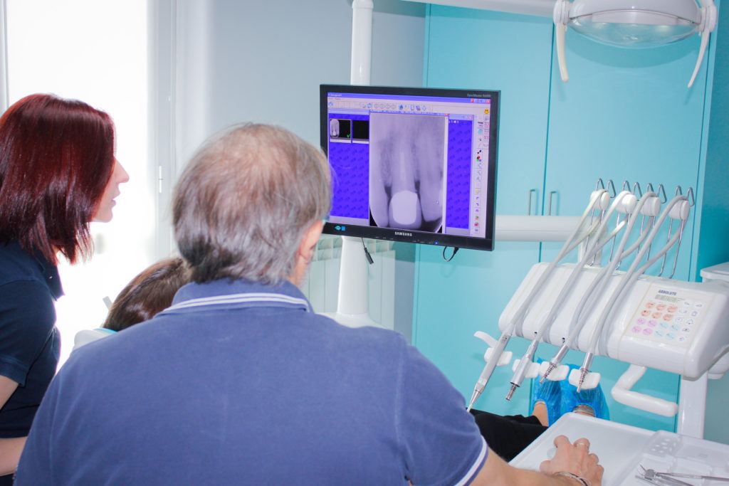
COMPUTED DIGITAL RADIOGRAPHY
The digital equipment of the latest generation ensures pictures of the highest quality and low exposure to ionizing radiation. It is no longer necessary to use X-ray films and chemicals for performing a radiographic examination, because the image is captured directly on the computer, providing excellent quality and high acquisition speed. You also have the ability to process and optimize the picture on your computer in order to avoid repeating the test if the result was not satisfactory.
Intraoral radiography
Panoramic radiography (OPG Ortopantomography) is not always enough to get a definitive diagnosis. For this reason it is useful to perform an intraoral radiography, i.e. a small x-ray done with the dentist’s X-ray machine, which allows to view up to three or four teeth the most. It is useful for example, during a root canal therapy, to identify the number and shape of the roots and to accurately measure the length of the canals. Furthermore, when starting a periodontal therapy, it is useful to have an intraoral radiography of all the teeth, in order to assess the amount of bone that is lost and the shape of the intraosseous pockets. The intraoral radiography is also useful when checking the presence of carious lesions that are difficult to view and it is also an important means of verifying the performed procedures (e.g. checks after implant treatment).
Implant planning using dedicated software
The implantologist has various diagnostic tools available. With the help of dedicated software, the surgeon receives the digital images on CD (in DICOM format) and thanks to them he can select the interesting anatomical areas. The program allows to virtually insert the implants in the required bone areas, being able to calculate their length and diameter and then perform the surgery with maximum safety and predictability. All this information can be transferred to another machine that quickly reproduces the surgical guides and crowns to be placed on the just inserted implants. It also allows you to make a virtual endoscopy – an instrument with major diagnostic value – for example, of the internal state of a maxillary sinus or the mandibular canal.
Intraoral photography
The intraoral photography has become an integral part of the complete treatment documentation. Thanks to the transition from analogue to digital, new procedures and imaging techniques have been developed that help the dentist to complete the diagnosis and document the progress and the final result of the therapy, as well as being a great help in communicating with their patients.
Software dedicated to motivating the patient
These are software packages that facilitate communication between doctor and patient, taking advantage of the effectiveness of video and demonstrative images that clearly and simply explain the established and proposed procedures, thus making the patient aware of all the treatment’s aspects that he will undergo, involving and guiding him with greater awareness towards the most appropriate choice.

